Our brain owns two blood supply systems: The Carotid arteries and vertebral arteries.
Carotid arteries
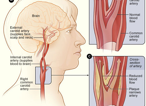
We can find the Carotid arteries on either side of the neck: Right and left. Figure 1 depicts only the right side set of arteries.
As we can see, the right carotid artery divides into two smaller branches: Internal and external. The same happens on the left side as well (A and B of this illustration).
The outer external branch transports oxygen and food to the face and all over the skull; the inner branch transports into the brain surface and inside structures.
Narrowing the route
Inset C depicts the narrowing of the route.
The junction where the common carotid artery divides into two is a crucial place. It is a common place where fat gets deposited and convert into a plaque.

Figure 1: Brain blood supply via carotid arteries
(Image: Wikimedia Commons, public domain)
Doctors can detect these narrowings. When it occurs, they can hear a sound, a “bruit”, with the stethoscope. However, when these narrowings are detected earlier, they can be removed by surgery as shown in Figure 2 below.

Figure 2: How carotid artery passage gets blocked and how it is removed
(Image: Wikimedia Commons, public domain)
Why it is so important?
They are at high risk of getting a stroke either by cutting off the blood supply or by dislodging plaque particles.
The plaque particles can travel higher through blood further inside and may cut off blood supply to a smaller area of the brain. This type of stroke, which is the commonest type, is called an ischemic stroke.
Blood supply to the base of the brain
Figure 3 (below) illustrates the blood supply on the skull base.
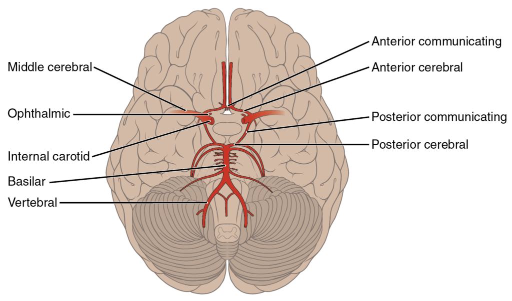
Figure 3: Blood supply routes on the base of the brain:
Source: openstax.org, RICE University under the CC BY 4.0 license
Different arteries to different brain regions
Different branches carry oxygen and nutrients to different regions of the brain’s surface. This is crucial. Figure 4 depicts these regions; the anterior cerebral artery carries supplies to the frontal lobe while the middle to the temporal and the posterior one to the occipital lobe.
Knowing these supply areas is very useful because the impairments due to an interruption to the blood supply in a stroke depend on which supply route is blocked.
For example, if the middle cerebral branch is blocked, the temporal lobe functions will stop.

Figure 4: Brain’s main blood supply areas according to its supply routes
Source: Frank Gaillard. Patrick J. Lynch, medical illustrator (Brain_stem_normal_human.svg) CC BY-SA 3.0
