Author: Prasantha De Silva
Module 7: Physical activities and exercise in stroke recovery
At the end of this module, you will be able to;
5.1. Describe the importance of initiating physical activity and exercise in stroke recovery
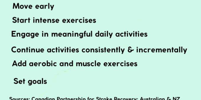
5.2. Principles of post-stroke exercises
5.3. Type of exercises we should promote and its frequency
5.4. Motivators and barriers to continue exercises
5.1. The importance of initiating physical activities and exercise as early as possible
In Module 4, we saw the brain’s recovery attempts after a stroke. We can help the brain in its rewiring process by initiating physical activities as early as possible. Physical activities bring a multitude of benefits not only moving arms and legs. The main advantages of early physical activities can be appreciated by reading this post: Detrimental effects of prolonged bed rest.
The main disadvantages of prolonged bed rest are as follows (source: The statement from the American Heart Association and the American Stroke Association).
- Losing muscle strength quickly
- Increased sodium and potassium loss through increased amounts of urine
- Decreasing immunity
- Losing heart muscle strength
- Increased risk of difficulty in moving limbs
- The difficulty of sitting and standing
Moreover, we cannot ignore that the physical activity also boosts self-confidence and psycho-social wellbeing.
The difference between the physical activities and exercise
We need to acknowledge the difference between physical activity and exercise at the very beginning. The term, physical activity, refers to any movement of the body and limbs that result in energy expenditure. The word, exercise, refers to a subset of physical activities that are planned, structured, and repetitive and are done with the aim of improving physical fitness (The American Heart and Stroke Association).
5.2. Principles of post-stroke physical activities and exercise
Research shows that we need to adhere the following principles for the maximum results.
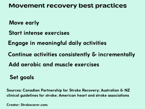
- Early mobility
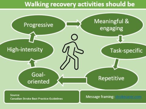
- Consistency of physical activities and exercise should be consistent and repetitive
- Repetitiveness
Early mobility: How early?
The American Heart Association and the American Stroke Association says the affected individual if they do not have medical or physical contraindications as certified by the physician, needs to sit on the bed within 24 hours after a stroke. This is because early mobility improves brain changes. You can read more about this topic here: “Early mobility improves recovery after stroke”.
Consistency and repetitiveness
- According to the American Heart Association and the American Stroke Association, physical activities is only effective if it is done consistently. This has been recognized as a formidable challenge for stroke survivors and stroke carers.
- Repetitive practice is the key: The US post-stroke rehab fact sheet equates this to a type of practice when one should do when playing the piano or pitching a baseball.
- The intensity of the exercises;
Exercise after stroke: http://hwcdn.libsyn.com/p/b/a/3/ba31d6360e144222/Zzz2.pdf?c_id=72611270&cs_id=72611270&expiration=1600313974&hwt=7ddf8d0963b4ee002d822d6d207dbf2f
exercises recommendations rational; https://www.ahajournals.org/doi/pdf/10.1161/STR.0000000000000022
5.3. Types of physical activities and exercise we should promote
Pre-requisites
Before embarking on an exercise program, a qualified health professional should screen and prescribe a suitable exercising program. This would be a challenge.
The Heart and Stroke Foundation outlines four types of exercises for stroke recovery. Those are as follows;
- Endurance (aerobic)
- Strength
- Balance
- Stretching
Exercises after stroke: An excellent but simple resource guide from the Stroke Foundation, New Zealand
It is rare to find online materials without copyright. However, I found an excellent open-access resource published by Margot Andrew, Margaret Hoessly, and Kate Hedges. It is under the Creative Commons Attribution Non-Commercial License (https://creativecommons.org/licenses/by-nc/3.0/), which permits unrestricted non-commercial use, distribution, and reproduction in any medium, provided the original author and source are credited.
Physical activities and exercise, and wellness videos for people with mobility challenges
A team of physiotherapists and occupational therapists from the University Health Network have published a list of exercises and wellness videos for people with mobility challenges. It includes an excellent collection of YouTube video clips. Here are some relevant ones. One should keep in mind that these resources do not replace the exercise regimen prescribed by your healthcare professional.
Three-part series of the “it’s Your Choice” project:
- Part I: An intro and disclaimer: https://www.youtube.com/watch?v=kpTHDR5k-cY
- Part II: The warm-up: https://www.youtube.com/watch?v=XPmUqiTBEpU
- Part III: Strength, balance, and aerobic exercises: https://www.youtube.com/watch?v=Qh94WI9Ecoc
Video exercises for those living with weaker one side
- Easy leg exercises: https://www.youtube.com/watch?v=-rwby0zA6Vs
- Hand exercises: https://www.youtube.com/watch?v=i0JYsLyJEnE
- Core exercises: https://www.youtube.com/watch?v=dGBqTLtdVuA
- Arm exercises: https://www.youtube.com/watch?v=kuuGlz_ddOM
How frequently one should do the rehab exercises?
How frequently one should engage in rehab exercises to gain a desirable effect? Janice Eng, a UBC physiotherapist has responded to this question. More specifically, the question was this:
How many upper limb reps per day can positively change the brain after stroke?
Quoting findings from other researchers (Nudo et al. 1996; Murata et al. 2007), she said it would be between 600- 1000 successful reach and grasp repetitions per day. The successful means if there are droppings it would not count. These findings were based on the research work of monkey brains after a stroke lesion to the motor cortex. The research has shown the hand representation in the undamaged primary motor cortex has regained. That is pretty impressive. The improvements have even progressed to precision finger grips.
To useful regain lost hand function, one should do at least more than 500 repetitive tasks a day.
Does this happen even in the best rehab centers? you can guess. It is not. Not even close to it. Catherine Lang and her team found it was about 32 per session (day). Later research that analyzed metadata showed the more the better.
5.4.Motivators and barriers to physical activities and exercise
Once started, the biggest barrier to better results is to maintain consistency. The barriers can be grouped into patient-related and environmental barriers, and lack of resources. The American Heart and Stroke associations detail out these barriers.
Barriers
Person-related | Context-related |
Depression | Lack of family and other social support |
Fatigue | Lack of access to fitness equipment |
Lack of motivation | Lack of trained personnel |
Fear of falling and other adverse effects | Cost |
Lack of knowledge about the usefulness | Lack of transport facilities |
Lack of perceived self-efficacy |
Motivators
- Providing psychological and social support
- Accessibility to trained personnel
- Establishing group classes
- Desire to achieve life goals
A free screen reader for the visually impaired
A stroke may either weaken vision or completely shut it off. And, those who are aged may have lower vision due to age. This is another barrier to improve communication of the stroke survivors as well as those already visually impaired.
Because of this, they are unable to socialize with their friends, read news, learn or engage in compatible jobs.
What if they have a method that it reads the computer screen? These are “screen readers”.
These are available, but expensive.
How about if you have it free?
Yes, while I was searching for that kind of facility, I found one: nvaccess.
A free screen reader: nv (non-visual) desktop access
According to their website, in 2006, Michael Curran began to develop a screen reader that could be run on Windows. Later, James Teh joined him. These two blind men founded this non-profit organization: NVDA (non-visual desktop access. Now, volunteers from all over the world have translated this into more than 55 languages and won multiple awards.
This is open-source software. What does that mean? it means that anyone can download the code free and those with knowledge can improve it further for use.
Module 6: Post-stroke rehabilitation
Introduction to the Module 6
This module discusses the concept of post-stroke rehabilitation. This is where stroke carers have a big role to play. Here, we discuss when the post-stroke rehabilitation begins, what aspects it includes, and the current expert recommendations.
Learning objectives
At the end of this module, you will be able to describe the components of post-stroke rehabilitation.
What is post-stroke rehabilitation?
It has three goals:
- Re-learning the lost skills
- Managing existing problems
- Preventing new problems
When and how the rehabilitation begins?
The experts say it begins at the hospital as soon as the affected is medically stable. It could be as early as 48 hours of the event. The earliest rehab activity could be turning and moving arms and legs while still on the bed. And, with professional supervision, sitting on the bed, moving between the bed and a chair are the next steps. It also includes the activities of daily living such as dressing, undressing, walking, bathing, using the toilet. Usually, personal healthcare workers assist. This is the first stage of returning to independence.
Domains of stroke rehabilitation
As documented in a comprehensive review, stroke rehabilitation runs along a continuum; first focusing on the function of an affected body part (earlier considered as impaired), second on the activities that can be done with that body part as a whole person (earlier considered as a disability), and third the participation in the social context or in a life situation (earlier considered as a handicap).
Elements of post-stroke rehab
The goal of post-stroke rehab is not only physical; it encompasses cognitive, social, emotional, psychological, communication, and occupational.
Elements of rehab
- Medical care: To manage underlying diseases such as high blood pressure, diabetes, heart disease, etc., and symptom management and also preventing another stroke
- Re-learning activities of daily living: grooming, dressing, undressing, toiling, moving, walking, and preparing meals, etc.
- Exercises: To strengthen muscles, improve coordination and balance, regain range of motion,
- Communication: improving communication skills and introducing to communication aids
- Swallowing: This goes together with communication improvement
- Vocational: Introducing to alternative income sources
- Social: strengthening social networks
- Mental, cognitive, and emotional
The most important element in rehab
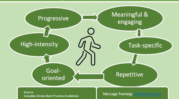
The National Institute of Neurological Disorders and Stroke says that the most important element in rehab is to create customized programs aimed at promoting repetitive activities that people engage in the mastering of “playing the piano or pitching a baseball”.
The most important element in rehab is to create customized programs aimed at promoting repetitive activities that people do when mastering “playing the piano or pitching a baseball”.
The national institute of neurological disorders and stroke
As we saw in Module 4, the brain “rewires” the alive neurons to carry out the tasks earlier carried out by dead neurons. We can speed up this by helping the affected individual to do repetitive exercises. Do you know that?
Researchers elaborate further on what repetitive exercises refer to.
Measuring outcomes and progress
For an individual who is undergoing the rehabilitation process is very important to know whether they are gaining anything. Over time researchers have developed a number of tools to monitor progress. A group of experts has grouped these measures under the three domains of rehabilitation cited above. Since it is a very comprehensive list, those interested can read about it through this link: http://www.ebrsr.com/sites/default/files/Chapter%2020_Outcome%20Measures.pdf
Module 5: Deficits and health problems after a stroke
Introduction
In Module 4, we discussed the brain’s recovery attempts after a stroke. However hard the brain attempts reverting to its pre-stroke functional level, it cannot regain everything lost; some of them will, however, reappear with time – mostly within the first six months post-stroke. Regaining the lost functions with time follows a non-linear trajectory as we learned in module 4. The rest will persist – as deficits – sometimes permanently.
And, new health problems such as pain and depression will also arise.
This module will look at the pre-stroke functions that may lose – called deficits hereafter – and the new health problems that are likely to develop. At the end of this module, you will be able to describe what those are and explain its relationship with the stroke attack.
The deficits directly related to the damaged brain locations.

5.1. Deficits that occur due to the Frontal lobe damage
When the ischemic stroke attacks occur due to a block in the middle cerebral artery, one of the commonest sites, cells of the motor homunculus dies. As a result, it will not be able to do its job; sending commands to muscles that move body parts starting from the eyelid, facial and mouth muscles, arm, and leg. As we now know that mostly, but not always, a stroke strikes one side of the brain, the deficit appears on the opposite side of the body. Depending on the extent of the damage, deficits can range from mild weakness to complete paralysis of one side of the body. Consequently, those affected will face the following difficulties.
- Movement difficulties
- Swallowing difficulties
- Speech difficulties
Movement difficulties
Movement problems are one of the most visible deficits that occur due to a stroke. The deficit could range from mild weakness to complete paralysis of one side of the body – from one side of the face, mouth, arm, or leg. For example, if the weakness is on the right side, the damage is on the left precentral gyrus in the brain and vice versa.
If the parietal lobe involves, the movement difficulties that may arise from the Frontal lobe damage, it can result in difficulties in maintaining posture and balance also. In a combination with these difficulties, walking becomes the greatest challenge.
Swallowing difficulties
This is also a common as well as serious problem since the affected individual can get choked with food.
Speech difficulties
The speech difficulties that occur due to the Frontal lobe damage will be discussed under 5.4. – language deficits.
5.2. Deficits that occur due to the Parietal lobe damage
Sensory disturbances
When the Parietal lobe’s post-central gyrus is affected, the affected person cannot feel touch, pain, as well as temperature. Following are the difficulties they face.
- Inability to feel touch, pain, and temperature
- inability to sense how the body is positioned
- Inability to recognize objects that they are holding
- Pain, numbness, and altered – tingling or tickling – sensation on the affected side, arm, or leg
- neuropathic pain – due to damage to the thalamus
- The heaviness of the affected arm or leg
5.3. Deficits that occur due to the Temporal lobe damage
The prominent deficit due to the Temporal lobe damage is to verbalizing thoughts through either speaking or writing. This occurs due to the damage in the Wernicke’s area of the lobe.
5.4. Language deficits (impairments)
Language deficits range from mild impairment to complete loss. The nature of deficit/s depends on the damaged area/s. The common language deficits are Broca’s aphasia, Wernicke’s aphasia, etc.
Broca’s aphasia
As the name itself implies, this language deficit results from the death of neurons in Broca’s area that situates on the Frontal lobe. It impairs expression either by speaking or writing; hence the name, “expressive aphasia”. However, if the Wernicke’s area is not affected, they can understand concepts but are unable to formulate grammatically correct sentences according to the National Institute of Neurological Disorders and Stroke.
Wernicke’s aphasia
As its name implies, this language deficit results from the death of neurons in the Wernicke’s area that situates in the Temporal lobe. In this type, those affected cannot understand the concepts, which others express either by writing or talking. Their communication is incoherent. The National Institute of Neurological Disorders and Stroke says that their expressions, although grammatically correct, have no meaning.
Speech problems
- Loss of expressing thoughts and feelings: they lose the ability to translate their thoughts and feelings into speech or writing language
- Difficulty in understanding spoken or written language and incoherent speech; they can make grammatically correct sentences but no meaning.
- Some can have the above difficulties together.
5.4. Mental, psychological, and cognitive deficits
After a stroke, a range of mental, psychological, and cognitive deficits may persist after initial recovery. It includes the following elements;
Difficulties in thinking and memory
- Deficits in short-term memory
- Difficulty in following instructions (apraxia)
- Loss of ability to respond to objects or sensory stimuli on the affected side
Emotional problems
- Fear, anxiety, sadness, anger, frustration
- Personality changes
5.5. Loss of bladder and bowel control
Another distressing problem is the loss of bladder control. However, the good news is that it can successfully be managed with proper knowledge and an easily acquired skill set. This will be discussed in a later module.
5.6. Pain
Researchers report pain as one of the commonest but under-recognized post-stroke problems. Those who live with a stroke report pain referring to any part of the body from head to toe.
Pain’s origin seems to be manifold; it can occur due to a stroke damage to the Parietal lobe, brain structures deep inside the brain – thalamus and pons, and due to “frozen” joints.
Pain as a result of “frozen” joints
The National Institute of Neurological Disorders and Stroke describes this pain occurs due to the non-use of joints for some time. This non-use leads to freezing tendons, ligaments, and muscles that assist in moving that particular joint.
Neuropathic pain
According to the experts in this field, those who experience this type of pain describe it as using these words: lacerating, aching, freezing, burning, and squeezing. They also may complain of pain even to just touching.
Although less common, this type of pain occurs as a result of the damage to structures deep inside the brain such as the thalamus and pons. Therefore, it is also called “thalamic pain syndrome”. Our sensory information from body parts to the brain transmit via the thalamus.
Spasticity-related pain
After a stroke, some develop spasticity in which the affected exhibit uncontrolled exaggerated movements. Experts say that two-third of them complain of pain and the majority of those who do not develop spasticity do not complain of pain.
Shoulder pain
Research reveals that shoulder pain is a common problem after stroke and may occur due to more than one reason including muscle weakness around the shoulder joint; however, pain most often appears with the development of spasticity. Prevention is the key. Experts advocate beginning physiotherapy “as soon as the patient is medically stable”.
This is only an introduction.
Please help us to fill the gaps in this post; add yours in the comments section.
Module 4: Brain’s post-stroke recovery attempts
At the end of this module, you will be able to describe;
- How our brain re-organizes after a stroke attack along a non-linear trajectory and adapts itself to continue its tasks
From the previous modules;
In Module 1, we discussed what the word, stroke, refers to: “Cell death that occurs in the brain, spinal cord, and retina attributable to the interruption to the blood supply”. It also dealt with stroke types including “mini-stroke” and characteristics of people at risk.
In Module 2, we discussed the brain basics: its covers, geography, regions and its assigned jobs, blood supply with relevance to the stroke.
In Module 3, we paid attention to stroke signs and symptoms, the “golden hour” concept, the F.AS.T. campaign, and how delays occur in seeking emergency care.
This Module 4 discusses how our brain fights back – its recovery attempts – when it comes under a stroke attack.
Non-linear trajectory of the brain’s recovery attempts
The brain’s recovery attempts follow a non-linear trajectory; it fights back faster in the first 3 months since the attack. Even from this period, the most improvements occur during the first few weeks. After that, it slows down towards the end of the sixth-month post-stroke.
Brain’s recovery attempts after a stroke attack follow a non-linear trajectory.
Most improvements occur in the first six months post-stroke.
However, there is good news; researchers have found the recovery attempts do not stop at the end of the sixth-month. It continues, although at a much slower pace, beyond the 12th month.
There is more good news: With appropriate training and other outside interventions, we can help the brain to speed up the process and achieve better recovery according to these researchers.
However, consider the non-linear trajectory as a general guideline.
We need to consider the above non-linear trajectory as a general guideline according to some researchers.
Why should we consider the non-linear trajectory as a general guideline?
This is because they have found that some patients recover faster and better than others. The trajectory seems to depend on the initial level of impairment and where the stroke occurs.
Figure 1 illustrates this phenomenon more clearly; Christian Grefkes and Gereon Fink, the authors of the article, Recovery from stroke: current concepts and future perspectives, published on June 16, 2020, in the Neurological Research and Practice journal, describes the graph: Those with milder impairment on admission recovers much better than those with severe impairment on admission.

Researchers say that recovery is strongly associated with the development of new connections within the affected areas. This post examines what those are and when these occur.
What happens within the first three weeks post-stroke
The brain attempts to restore supply routes.
As soon as a stroke attack unleashes, the brain begins to strike back. Its alarm system activates the alternate collateral blood supply mechanisms to sustain oxygen and food supply to neurons and its supportive cells. Most of the time, this back-up mechanism cannot save all the cells. Those who live inside the areas directly affected begin to die. This area is called the “infarct area” or the “ischemic core”. Meantime, its neighboring cells, become hyperactive, in spite of reduced supplies. This region is called the “penumbra”.
With time, if the brain becomes unable to sustain its blood supply above the critical threshold, the cells in the penumbra region also succumb adding to the infarct area; as a result, the infarct area becomes larger and larger. Figure 1 illustrates the scenario beautifully.

(source: Stroke: past, present, and future by Macrae M and Allen S.M.; Brain and Neuroscience Advances,
2018; SAGE open access publications under the CC-BY-NC 4.0.
Attempts to restoring functions
The hyperactive cells in the penumbra begin to release growth molecules. They stimulate cells to begin “rewiring” and as a result, new dendrites and axons sprout by the second and third weeks. And new synapses too. The aim of these attempts is to establish new connections with still-alive neighbors to resume lost functions earlier carried out by their dead neighboring cells (Jillard et al. (2005). However, some researchers say these rewiring efforts sometimes hinder the task of initiating new meaningful connections too.
What happens between the third week and the 6th month post-stroke
Tissue repair (“rewiring”)
By the third week, the brain establishes its rebuilding project through neural repair- “rewiring”. It includes sprouting axons, generating new neurons, and glial cells too.
The neighboring cells who survived from the attack acquire some of the functions earlier carried out by their now-dead neighbors. The National Institute of Neurological Disorders and Stroke says the practice helps in this “rewiring” process. We can accelerate the process by initiating an intense rehab program within the first week according to the researchers.
However, thee changes do not limit to the affected regions; the whole brain seems to make changes within its neural networks including in the non-affected half of the brain according to the researchers. And, these changes apply not only in recovering motor deficits but in language recovery too.
Module 3: Stroke warning signs and symptoms
Introduction to the module 3:
Module 3 discusses common stroke warning signs and symptoms, how those relate to the brain basics that we discussed in Module 2. Then, we will delve into the popular F.A.S.T. stroke awareness campaign. I encourage you to re-visit module 2 – brain basics – for a better understanding of how the warning signs occur.
Learning objectives:
3.1. Stroke warning signs and symptoms
3.2. The F.A.S.T. campaign
3.3. The concept of the “golden hour”
3.4. Delays in seeking hospital care
3.5. What should we do in those situations?
3.1. Stroke warning signs and symptoms
According to the National Institute of Neurological Disorders and Stroke1, the following are a list of stroke warning signs and symptoms.
- Sudden numbness or weakness of one side of the face, an arm, or a leg
- Sudden confusion
- Difficulty in understanding or trouble in speech
- Sudden trouble seeing in one or both eyes
- Sudden trouble walking, dizziness, loss of balance and coordination
- Sudden severe headache with no known cause
- Seeing two images (double vision)
- Vomiting
If you re-visit Module 2: The brain basics, you can appreciate how these problems occur as a result of interruption to the blood supply routes; anterior, middle, and posterior cerebral arteries.
As soon as the signs and symptoms begin to unfold, no one can be sure whether it is going to be either a stroke or mini-stroke. Hence the safest strategy in this scenario is to suspect a stroke and take the individual to the nearest hospital with a minimum delay. Even if it is a mini-stroke, the patient needs to be assessed as early as possible to prevent a full-blown stroke.
3.2. The F.A.S.T. campaign
F.A.S.T. is an acronym of popular stroke warning signs and symptoms awareness campaign in the world. Each of the first three letters denotes one common sign or a symptom of stroke, which can easily be detected by anyone. The last letter, T, emphasizes the urgency of taking action – call emergency – as soon as possible. Those are;
- F : face,
- A :arms,
- S : speech,
- T : time.
The following poster (Figure 1) is from such a campaign carried out by the University College London Hospitals in the NHS. It summarises what F.A.S.T. means.

How should we use the F.A.S.T.?
Knowing what these letters represent is not enough. We need to ask its associated specific questions. The following questions assist you in the effective use of the acronym.
How to suspect a stroke
- Look at the face
Ask: can you smile?
Observe: whether one side of the mouth or an eye is drooping - Compare both arms
Ask: Raise your both arms
Observe: Whether the person is having any difficulty of raising one or both arms - Observe the speech
Observe: whether the person cannot speak or understand as before
- Check the time
Call an ambulance of you observe any one of the above
However, research reveals that the F.A.S.T. campaign, although it raises the awareness of stroke signs and symptoms, may not significantly improve our response to the situation, particularly when the signs and symptoms are not severe such as in the case of mini-stroke2.
3.3. The concept of the “golden hour”
In the event of a stroke, the first hour is the most critical to save brain cells as many as possible. As you already know every passing minute costa about 2 million neurons3. The affected person should be on the bed of a hospital with adequate facilities to manage a stroke emergency. This hour is described as the golden hour4.
However, the elapse of the first hour does not mean that we should lose our hope of salvaging still-alive but affected brain tissues. The Canadian best practice guidelines keep from the “witnessed symptom onset” to hospital arrival time as “four and a half hours”5.
The following video clip from the US CDC summarizes the stroke signs and symptoms except for its reference to the time frame.
3.4. Delays in seeking hospital care
Saving still-alive brain cells is a race against time. However, delays happen. The delays can be conceptualized as pre-hospital delays and in-hospital delays. This section discusses the reasons for prehospital delays.
3.4.1. The “family member” effect
Interestingly, research shows that if a stroke occurs at home and in front of family members and loved ones, the delaying time to seek hospital care is longer than if it occurs at the workplace or in front of unknown bystanders6.
This is important because the majority of strokes occur at home and they arrive late at the hospital. For example, in the US, 70 percent of stroke events occur at home and 70 percent of patients with a suspected stroke arrive at a hospital six hours after the event6.
3.4.2. Knowledge and perceived seriousness
Lack of knowledge about stroke warning signs and symptoms is a major problem in spite of awareness campaigns. This is especially relevant to low-middle income countries where the majority of strokes occur. Very few studies exist about awareness levels of stroke warning signs and symptoms from those countries.
However, the knowledge of warning signs alone is inadequate to shorten the delay. It is not the knowledge but, research shows, the perceived seriousness of the observed signs and symptoms triggers action8.
3.4.3. Communicating suspected stroke signs and symptoms
The delay also occurs after taking the decision either to call an ambulance or taking to a hospital depending on the services available. This occurs when describing the event via telephone because most of the time people use vague descriptions. Most of the time we tend to use vague terms to describe the events of a stroke9.
3.4.4. Transport
Lack of transport facilities is a major cause for delay in places where community ambulance services either do not exist or not accessible due to geographic and economic reasons, particularly in low-middle income countries.
3.5. What to do when we encounter a suspected patient
- Decision making is the key; Follow the F.A.S.T.
- First aid and CPR if necessary
- Remind the reasons for the delays
- Act fast; call an ambulance; if that facility does not exist, take the person to the nearest hospital as soon as possible.
References
- National Institute of Neurological Disorders and Stroke (NINDS): Basic facts: preventing stroke; NIH; 2020. Accessed on September 16, 2020.
- Wolters FJ, Li L, Gutnikov SA, Mehta Z, Rothwell PM. Medical Attention Seeking After Transient Ischemic Attack and Minor Stroke Before and After the UK Face, Arm, Speech, Time (FAST) Public Education Campaign: Results From the Oxford Vascular Study. JAMA Neurol. 2018;75(10):1225–1233. doi:10.1001/jamaneurol.2018.1603
- Jeffrey L. Saver (2006): “Time is Brain”: Quantified; Stroke Journal. 2006;37(1): 263-266.
- Advani R, Naess H. & Kurz M.W. (2017). The golden hour of acute ischemic stroke.Scand J Trauma Resusc Emerg Med. 2017 May 22;25(1):54. doi: 10.1186/s13049-017-0398-5.
- Canadian Stroke Best Practices (2018): Emergency Medical Services Management of Acute Stroke Patients Recommendations.
- Dhand, A., Luke, D., Lang, C. et al. Social networks and risk of delayed hospital arrival after acute stroke. Nat Commun 10, 1206 (2019). https://doi.org/10.1038/s41467-019-09073-5.
- Eric S. Donkor (2018): Stroke in the 21st Century; Stroke Res Treat.: published online.
- Teusch I Y, Brainin M. Stroke Education: Discrepancies among Factors Influencing Prehospital Delay and Stroke Knowledge. International Journal of Stroke. 2010;5(3):187-208. doi:10.1111/j.1747-4949.2010.00428.x
- Christopher T. Richards, Baiyang Wang, Eddie Markul, Frank Albarran, Doreen Rottman, Neelum T. Aggarwal, Patricia Lindeman, Leslee Stein-Spencer, Joseph M. Weber, Kenneth S. Pearlman, Katie L. Tataris, Jane L. Holl, Diego Klabjan & Shyam Prabhakaran (2017) Identifying Key Words in 9-1-1 Calls for Stroke: A Mixed Methods Approach, Prehospital Emergency Care, 21:6, 761-766, DOI: 10.1080/10903127.2017.1332124
Module 2: Brain Basics for stroke carers
At the end of this module – brain basics for stroke carers – you will be able to describe the brain’s basic anatomy and functions that could be affected by stroke.
In Module 1 – Stroke basics for stroke carers – we peeped into the definitions of the word, stroke, different stroke types, what happens in a stroke, and who are at risk.
2.1. Brain covers
2.2. Brain surface map
2.2.1. Frontal lobe and Broca’s area
2.2.2. Parietal lobe
2.2.3. The “Homunculus” (“Two Little Humans”)
2.2.4. Temporal lobe
2.2.5. Occipital lobe
2.3. Brain’s blood supply
2.1. Brain covers
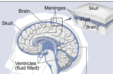
Brain basics for stroke carers begin with an introduction to brain covers. Our brain is a very pliable organ. Even a small finger pressure makes a dent over its surface. The bony skull protects it and, as Figure 1 shows, three layers underneath the skull wrap the brain snugly. The outermost cover ( the folded one in Figure 1) is called, “Dura mater” (meaning “tough mother”). As its name implies it is firm and thick. The other two inner covers (“spider mother” and “soft mother”) is thinner, transparent, as well as glistening. Blood vessels traverse through the two layers bathing a colorless “cerebrospinal fluid”. You can also see that the surface is folded into grooves (long convoluted ditches) and bumps (long convoluted surfaces) underneath the inner two covers.

Visual credit: Videos drawn from the NeuroLogic Exam and PediNeuroLogic Exam websites are used by permission of Paul D. Larsen, M.D., University of Nebraska Medical Center and Suzanne S. Stensaas, Ph.D., University of Utah School of Medicine. Additional materials were drawn from resources provided by Alejandro Stern, Stern Foundation, Buenos Aires, Argentina; Kathleen Digre, M.D., University of Utah; and Daniel Jacobson, M.D., Marshfield Clinic, Wisconsin. The movies are licensed under a Creative Commons Attribution-non-commercial-ShareAlike License.
2.2. Surface map
Our brain surface owns a highly advanced map. Let us see how it is organized. An understanding of this map is an essential part of brain basics.
Figure 2 shows the brain sans its covers. It consists of two equal halves – identified as left and right hemispheres. Both communicate with each other via neurons cells who travel through a short thick stalk deep in the middle. We do not see the stalk in this image.

Scientists name this densely wrinkled (or densely folded) surface as the “cerebral cortex”; it is a layer with thickness varying from 1mm to 4.5mm averaging about 2.5mm (one inch)1. This layer, grey in color (also called “grey matter”) is made up of tightly packed millions of neurons. Using its grooves and bumps as landmarks, scientists divide it into four large regions, also called “lobes”: Frontal lobe, Parietal lobe, Temporal lobe, and Occipital lobe (Figure 3).

2.2.1. Frontal lobe (“region”)
Figure 3 depicts the Frontal lobe blue in color. It extends front to back until it meets a groove (or a fissure), scientists name the”Central Sulcus”. This groove separates the Frontal lobe from another two regions (lobes): the Parietal lobe and the Temporal lobe.
Pre-central gyrus (Primary Motor Cortex)
Now, look at Figure 4 and focus on the red-colored strip that lies in front of the Central Sulcus. Scientists name this bump the “Precentral Gyrus” because of its placement. As you can see, this strip ascends parallel to the Central groove from its left side over to the top. It ends after descending about 1cm on its own. We have exactly a similar strip on the right half of the brain too. The nerve cells that populate this strip has a very special job to do; they send commands, with our approval, to muscles of the opposite side of the body to move. Because of this job, scientists call it the “Primary Motor Cortex”.

(Image courtesy: S Bhimji MD; from Neuroanatomy, Postcentral Gyrus Copyright © 2020, StatPearls Publishing LLC. under the terms of the Creative Commons Attribution 4.0 International License (http://creativecommons.org/licenses/by/4.0/),
The nerve cells in the large grey-colored area that spans towards the front of this “Primary Motor Cortex” become very busy when we think, make decisions, behave in ways whatever we want. Because we, humans, are the only ones who can do these high-end activities, we own the largest Frontal lobe among all animals.
Broca’s area (Figure 5)
This is a special small area in the Frontal lobe that merits our attention. We find it over the left Frontal lobe. The nerve cells in this area specialize in word production both in speech and writing. They process information that receives from another area (Wernicke’s area that situates in the Temporal lobe) and coordinates those with another set of large neural networks2.
Why do we need to know about this? It is because the death of these nerve cells results in a special kind of speech difficulty, called Broca’s aphasia.

(source: Wikimedia Commons)
2.2.2. Parietal lobe (Figures 3 and 4)
Figure 3 depicts this lobe (“region”) pale white in color. It begins just behind the Central Sulcus (groove), spreads over the top, and descends towards the left side to meet another groove, called the Lateral Sulcus. And the lobe extends behind to meet another groove that separates it from the Occipital lobe.
Post-central Gyrus
Figure 4 depicts this area of strip blue in color. Beginning just behind the Central Sulcus (groove), it runs parallel to its counterpart, the Precentral Gyrus that lies on the opposite bank of the groove. Very much similar to its counterpart, the nerve cells in this area look after the exact same locations of the body’s opposite side. However, there is a marked contrast; unlike its counterpart nerve cells who send commands, these nerve cells receive information about touch, temperature, and pain. Then, they relay the information to the Frontal lobe nerve cells for necessary action. Because of the nature of this job, scientists name this strip the “somatosensory cortex”. For example, when we feel pain in the left arm, the area assigned in this strip receives the information and then relays the information to the Frontal lobe. After processing this information, the nerve cells in the “primary motor cortex” send instructions to move the left arm from the source of the pain.
The other areas of the parietal lobe carry out more complex jobs than the above, however. It includes maintaining balance, recognizing objects, etc.
2.2.3. The two Homunculi (“two little humans”) (Figure 6 and 7)
This is a very useful excellent metaphor with practical value to understand the brain basics for stroke carers. In Figure 4, we found red-colored and blue-colored strips on either side of the Central Sulcus (groove). While the nerve cells of the former are responsible for sending commands to move muscles, the nerve cells of the latter strip process sensory information from the body.
Researchers have shown that both strips own a unique layout to execute their jobs. If we climb the strip (precentral gyrus) starting from the area closest to the left Lateral Sulcus (groove) (Look at Figure 4) until it ends at the other side, we will find a homunculus; a small replica of the human body. Compare Figures 6 and 7.
The layout is exactly similar to a homunculus: a small replica of a human body. The “Homunculus” refers to a fictional fully formed human being but extremely small in size; it is a miniature replica. Figures 6 and 7 re-create it beautifully. We own two homunculi on each side of the brain: The “motor homunculus – the strip that lies in front of the Central groove, and the “sensory” homunculus that lies just behind the Central groove. Another interesting characteristic is that the assigned area’s sizes are proportional to the complexity of the job to do. For example, the assigned areas for the fingers, tongue, and face are much larger than the rest of the areas.

(Source: Wikimedia Commons under the CC BY 3.0 license)
(Source: Wikimedia Commons under the CC BY 2.1 Japan license)
Dr. Wilder Penfield and Edwin Boldrey in 1937 “discovered” and wrote a detailed 50-page article with drawings.
We already know what these “little humans” are doing there.
2.2.4. Temporal lobe (Figure 3 and 4)
Figure 3 depicts the left Temporal lobe (region) green in color. It extends from the “Lateral groove” (also called “Lateral Fissure”) back until it reaches the Occipital lobe’s territory. The nerve cells in this region process information about language understanding, hearing, and memory.
Wernicke’s area
The neurons in this special area process information of language understanding and interpretation. Then it relays the information to the Broca’s area’s neurons for word production.
When we hear something, the hearing area receives information from the ear and then send this information to the Wernicke’s area. Similarly, when we see or read something, the visual area receives it and then sends that information to the Wernicke’s area. Neurons in this area work hard to retrieve suitable nouns appropriate to the context from the storeroom, set the language structure, and shoot the processed information to the Broca’s area.
The death of brain cells of the Wernicke’s area will result in a specific language disability called “Wernicke’s aphasia”. We will learn about it in another module.
2.2.5. Occipital lobe (Figure 3)
This lobe appears colored brown in Figure 3 and situates the brain’s back surface. Its neurons interpret images that receive from our eyes. The damage to neurons in this area causes loss of our sight even though the eyes and its nerves are intact.
The above layout of brain basics for stroke carers provides a solid foundation to improve care for those living with a stroke.
2.3. Brain’s blood supply
Another useful area of the brain basics for stroke carers are the brain’s blood supply architecture. The brain receives oxygen and food for survival via two main supply routes: carotid arteries and vertebral arteries. The carotid arteries climb from the sides of the neck while the vertebral arteries climb through our vertebral column which situates the back of the neck. Inside the brain, they form an inter-connected-circle, called the circle of Willis.
Carotid arteries
Figure 8 still-image taken from a video clip from a demonstration-explanation of the brain’s blood supply by a professor of the University of Utah shows the Left side carotid artery and its divide. This is the main supply route. We own a similar one on our right side too.

At the level of the neck, the carotid arteries, both left and right, branch out into two: External and internal. The latter again divides into three smaller ones: anterior (front), middle, and posterior (back).
Figure 9 illustrates their feeding areas in the brain; the first one carries blood to the neurons who manage information related to the lower part of the body including the legs; the second one (the middle branch) nourishes the neurons who manage information related to the upper part of the body including the face, tongue, arm. This also includes areas assigned to language and speech: Broca’s area and Wernicke’s area.

Frank Gaillard. Patrick J. Lynch, medical illustrator (Brain_stem_normal_human.svg) CC BY-SA 3.0
Why should we know about this information? This knowledge of brain basics for stroke carers is critical to treat and providing care to the affected. For example, if the right middle branch’s blood supply interrupts, the affected person will show difficulties in raising or feeling of the left arm and drooping of the right side of the face, etc. If the left middle branch’s blood supply interrupts, in addition to the above, we can observe speech and understanding difficulties also.
References
- Bruce Fischl, Anders M. Dale (2000): Measuring the thickness of the human cerebral cortex from magnetic resonance images bruce Fischl, Anders M. DaleProceedings of the National Academy of Sciences Sep 2000, 97 (20) 11050-11055; DOI: 10.1073/pnas.200033797.
- Redefining the role of Broca’s area in speech Adeen Flinker, Anna Korzeniewska, Avgusta Y. Shestyuk, Piotr J. Franaszczuk, Nina F. Dronkers, Robert T. Knight, Nathan E. Crone Proceedings of the National Academy of Sciences Mar 2015, 112 (9) 2871-2875.
Module 1: Stroke basics for stroke carers
Module 1 for stroke carers introduces what the word – stroke – refers to, what happens in a stroke event, types of stroke, and who are at risk.
Learning objectives:
At the end of this module, you will be able to answer the following questions if someone asks.
1.1. What is a stroke?
1.2. What happens in a stroke?
1.3. How does a stroke occur?: Types of stroke
1.4. What is a “mini-stroke”?
1.5. Who are at risk?
1.1. What is a stroke?
The term, “stroke” is a broad one; a more recent definition from the American Heart Association/ Stroke Association advocates use this term to cover cell death that occurs in the brain, spinal cord, and retina attributable to the interruption to the blood supply. According to this definition, the presence of symptoms or signs of more than 24 hours is not essential to use the term. And, on the other hand, the presence of reversible stroke-like symptoms more than 24 hours due to edema without interruption to the blood supply does not qualify for a stroke.
However, “Stroke” is a disease of the brain, not of the heart. Many believe, incorrectly, that it is a type of heart disease. Since it occurs suddenly, some call it a “brain attack”. Health professionals name the condition as “cerebrovascular disease”.
Stroke is a brain disease,
not a heart disease.
Stroke is the second leading cause of death and the third leading cause of disability in the world. As much as 70 percent of stroke events are thought to occur in low-middle income countries. On average, a stroke occurs 15 years earlier among people living in those countries. During the past four decades, stroke incidence among low-middle income countries has doubled while it has halved in high-income countries1.
A stroke occurs as a result of brain cell death due to an interruption of blood supply to a part/s of the brain. We will discuss this in module 2.
Stroke warning signs are easy to detect. The common ones are drooping one eyelid, inability or difficulty of raising one or both arms, and slurred speech; there are more. We will discuss these in module 3.
However, a stroke can also occur without any observable changes.
Stroke results in a range of disabilities of the affected person and at least half of these disabilities persist permanently for life, if not treated early. It exerts an enormous burden not only on the person affected but the whole family, and the society. We will discuss various kinds of disabilities in detail and what we can do to rehabilitate those in Part II of this course.
1.2. What happens in a stroke?
A stroke cuts off blood supply to a part of the brain. (You can learn more about the brain by joining the “journeys to the brain” series). Within minutes, the neuron cells in the affected area begin to die at a rate of 32,000 per each passing second. In terms of minutes, the number amounts to about two million neuron cells per minute2.
Stroke kills about 32,000 neurons in each passing second.
Jeffrey L. Saver (2006): “Time is Brain”: Quantified; Stroke Journal. 2006;37(1): 263-266.(message creator:https://www.strokecarer.com/)
The only way to save the rest of the cells threatened with death is to restore the interrupted blood supply as soon as possible. This can only be done in a hospital with adequate facilities.
1.3. Types of stroke
A stroke usually occurs either due to a block in a supply route or a burst of the supply route’s wall.
Stroke can occur due to a block inside an artery to the brain or a burst of its wall.
1.3.1. Ischemic stroke
Ischemic stroke occurs due to a block by a blood clot inside the supply route. The extent of the damage depends on where the clot clogs the blood supply system inside the brain. If a clot is a bigger one, it clogs a larger artery and blocks the blood supply to a larger area of the brain. If the clot is a smaller one, it may travel as far higher up as possible until it clogs a smaller branch of a smaller artery.

How a blood clot forms
A roughened wall of an artery triggers blood clot formation. The roughening begins with fat deposition at one place of the wall. Then, it hardens with calcium and cholesterol deposits. The process continues slowly but surely forming a plaque there. This thickening narrows the lumen and roughens the surface. Plaque build-up can occur anywhere; however, strokes are commonly associated with plaque build-up in the neck vessels – carotid arteries as shown in Figure 1.
The following video clip from the British Heart Foundation explains simply how arterial thickening occurs and the factors that facilitate the process.
A clot can even originate inside the heart particularly among those with heart problems and travel into the brain.
In the US, about 80 percent of all strokes are in this nature according to the National Institute of Neurological Disorders and Stroke3. In low-middle income countries, this percentage is about 66 percent1.
1.3.2. Hemorrhagic stroke
When a stroke occurs due to a burst of the vessel wall, we call it a “hemorrhagic stroke” (Figure 2). As a result, blood seeps out of the vessel, and oxygen and food supply to neurons interrupt. The bleeding exerts pressure on the area causing more damage.

However, there is another 5-10 percent of people who develop a stroke due to an unknown reason4.
1.4. What is a “mini-stroke” TIA (Transient Ischemic Attack)?
In this situation, the stroke signs and symptoms last less than 24 hours; most often, less than an hour. Hence, it is also called “Transient Ischemic Attack” (TIA). What happens here is that the clot that blocks the supply route disappears after a brief time.
However, it is a dire warning; it will certainly return as a full-blown stroke, often within the first week after the TIA, if not treated5.
Therefore, a mini-stroke should also be considered as a medical emergency.
In the following video clip, Professor Peter Rothwell explains why we should a mini-stroke also as a medical emergency.
However, if the effects last more than 24 hours, it is considered a stroke. Ideally, anyone who experience a mini-stroke should not drive or operate a machinery for a month. In some countries it is the law.
Do not drive after a mini-stroke at least for a month. In some countries, it is the law.
1.5. Who are at high-risk?
Some are at higher risk for stroke. We can reduce the risk by modifying some risk factors while others are non-modifiable. The source of the following list is the National Institute of Neurological Disorders and Stroke3.
Stroke can strike even the very young; no one is immune.
Non-modifiable risk factors
- Age: The risk doubles every passing decade from 55 to 85. However, it can also occur in childhood6.
- Sex: Men are at higher risk in young and middle age. In older ages, however, the risk is equal.
- Ethnicity: some ethnic groups are at higher risk; for example, African-Americans and Hispanics experience stroke events more than Caucasians.
- Family history
Modifiable risk factors
There are some risk factors that we can reduce the risk. Those are as follows;
- High Blood Pressure: This is one of the most potent risk factors we can easily modify.
- Diabetes
- High cholesterol
- Cigarette smoking: This raises the ischemic stroke risk by two-fold and hemorrhagic stroke risk by four-fold.
- Physical inactivity and obesity
References
- Johnson, W., Onuma, O., Owolabi, M. & Sachdev, S. Stroke (2016): a global response is needed. Bulletin of the World Health Organization 94, 634–634A, https://doi.org/10.2471/BLT.16.181636.
- Jeffrey L. Saver (2006): “Time is Brain”: Quantified; Stroke Journal. 2006;37(1): 263-266.
- National Institute of Neurological Disorders and Stroke (NINDS): Basic facts: preventing stroke; NIH; 2020. Accessed on September 16, 2020.
- Donkor E.S. (2018): Stroke in the 21st Century; Stroke Res Treat.: published online.
- British Medical Best Practice; accessed on September 20, 2020.
- Vrudhula A, Zhao J, Liu RToo Young to Have a Stroke?—a Global Health CrisisStroke and Vascular Neurology 2019;4:doi: 10.1136/svn-2019-00029.
Detrimental effects of prolonged bed rest
Have you thought about this? About the detrimental effects of prolonged bed rest? This post is about that.
A little bit of history about bed rest research
Researchers say that bed rest was considered as a treatment strategy in the 19th century. This view was beginning to change at the turning to the 20th century.
In 1947, Dr. R.A.J. Asher wrote an article to the British Medical Journal about “Dangers of going to bed”. His article was meant for physicians: ” we should think twice before ordering our patients to bed and realize that beneath the comfort of blanket there lurks a host of formidable dangers”.
Since then, more evidence has been accumulating. In 1999, a group of researchers reviewed 39-bed rest trials published in The Lancet. The conclusion: bed rest did not improve the outcome significantly; rather they reported nine situations with worsening outcomes.
However, the campaign is ongoing. For example, if you visit the website of the American Academy of Nursing, you can read the “Don’t Statement”:
Don’t Statement
Don’t let older adults lay in bed or only get up to a chair during their hospital stay. Walking during the hospital stay is critical for maintaining functional ability in older adults.
American Academy of Nursing
In fact, The problem has received attention from several countries. It has led to a campaign named, “End PJ paralysis”.
End PJ (Pyjama) paralysis
End PJ paralysis is a global movement: https://endpjparalysis.org/

The following is a poster that is aimed at addressing this problem. We can find a series of similar educational tools to reduce the detrimental effects of prolonged bed rest.

Let us dive into our bodies to find out what happens when we take prolonged bed rest.
What happens inside our body during a prolonged bed rest?
All our body mechanisms are set to function best when standing upright – and, to sleep only about eight hours. If we prolong our bed rest time for more than 24 hours, the body begins to re-set all the systems to face the new challenges. Certainly, it will. And, it will result in a series of detrimental effects of prolonged bed rest.
Let us jump into this journey of exploring the “detrimental effects of prolonged bed rest”.
Prolonged bed rest’s detrimental effects on the heart and our blood circulation
In an upright position, most of our blood circulates below the heart level. Veins bring up the returning blood with all waste products produced by cells including carbon dioxide. The valves in veins and muscles support veins to do the job.
In contrast, in a lying down position, blood slowly moves to the abdomen, lower back, and lungs from the legs. The new situation exerts pressure on the heart. To relieve the pressure, the body initiates mechanisms to remove a certain amount of water from our blood through kidneys. The aim is to reduce the burden – preload – on heart output. Not only that, but the prolonged bed rest also reduces red blood cell mass too to reduce our blood’s oxygen-carrying capacity.
If the bed rest continues as long as 6 weeks, research has shown that the heart muscles can get atrophied. If the bed rest continues for 20 days, the heart output can reduce by 25 percent according to Kristin J. Stumpfle and Daniel G. Drury.
These adjustments cause problems; one is to increase the resting heart rate; another is the postural hypotension in which we feel dizzy when we attempt to either sit on the bed or stand. It can occur even after 24 hours of strict bed rest.
Another interesting adjustment occurs in our venous blood collection system. It begins to pool blood at our deep veins. As a result, the risk of developing blood clots increases leading to deep vein thrombosis. And, the formed blood clots can dislodge, travel all the way up to lungs, and stuck there. This can result in pulmonary embolism, always a fatal situation.
Prolonged bed rest’s detrimental effects on our muscles
Very much similar to the heart and blood circulation mechanism, our muscles also work best when we stand upright against gravity. In a prolonged bed rest, with time, they begin to shorten and then remove some of its muscle fibers. It invariably loses muscle mass and subsequently its strength. Research shows that we can lose muscle strength by 6 – 40 percent within 4 – 6 weeks of complete bed rest. More recently, a group of researchers from Johns Hopkins found that each passing day in the ICU lowers muscle strength by 3- 11 percent a day over the ensuing months and may even extend to years.

As expected the most affected muscles are the ones that work against gravity”: The “anti-gravity” muscles. Those are the muscles that help to raise the foot at the ankle joint (plantar flexors), those in the thighs and arms (quadriceps and hamstrings), those in the buttocks, calves, lower back, abdomen, and the neck. In some muscles such as those in calves, thighs, and feet, we can readily see the wasting; however, in other muscles, we cannot readily see. Research shows that the process of wasting begins as early as on the fifth day and reaches its peak in the second week of bed rest.
Effects on joints
Muscles are attached to joints through tendons and ligaments. And, joints are covered by some cartilage. Because of non-use, fibers in tendons and ligaments become shortened. Surrounding connecting tissues turn rigid due to the addition of collagen. The result? the development of almost permanent contractures that freeze joint movements. Research shows that the appearance of collagen fiber can be observed as early as on the sixth day of complete bed rest.
These changes occur in all joints. But, it is most pronounced in the hip, knee, and ankle joints.
Effects on bones
As in every part of our body, bones also respond negatively to bed rest. It begins to weaken with time; its building block – calcium – starts appearing in our urine within a few days of bed rest. It also increases the risk of forming kidney stones and urine infections. To make matters worse, calcium absorption in the intestine also decreases.
Research reveals that the bones in our legs and lower back are the worst affected.
Effects on the kidneys and bladder
Due to non-use of bones, its building block, calcium, beings to drain into urine. During the process, the chances of forming stones inside kidneys and the bladder rises. Furthermore, due to urine rentioninside the bladder the chances of urine infection als rises.
Effects on the skin
The effects of prolonged bed rest on the skin particularly the skin over bony prominence are two-fold; shear and friction damage the superficial parts of the skin while the pressure interrupts the deep tissue functions. It includes underlying muscles too.
Prolonged bed rest due to the pressure it exerts on the skin over bony prominences occludes the smallest blood carriers – capillaries – blocking the blood supply to the skin and its surrounding tissues. This sudden attack deprives living cells of oxygen and nutrient supply. The situation will lead to cell death.
Experts say that the critical duration of pressure that requires developing a pressure injury can vary from 30 minutes to 4 hours. This variation depends on underlying diseases that affect small blood supply vessels including the smallest – capillaries.
Shearing, in addition to the direct external pressure, contributes to skin damage. Shearing refers to lateral displacement of the skin due to traction over the surface. Moreover, moisture too worsens the situation by softening the skin layers.
Not only the external pressure, but shearing and friction on the skin damage the skin also. It deprives the skin cells and underlying tissues of its oxygen and nutrition for their survival, It can result in devastating bedsore. Once the process sets in, it can become a slippery slope. The most common 5 sites that pressure ulcers occur are the heel, ankle, bony prominences over the sides of the hip, sacral area, and skin over the sitting bones in the buttocks.
In fact, the detrimental effects of prolonged bed rest manifest all over the body.
Glial cells: Journeys to the brain-14
“Neurons Converted from Glial Cells” by National Institutes of Health (NIH) is licensed under CC BY-NC 2.0
Can we create neurons from another cell type? Yes!. The red-colored cells you see in the above image are neurons that were originally mice glial cells. NIH researchers have converted those into neurons using gene therapy.
Furthermore, these new neurons, they have demonstrated, can do the lost neurons’ job!.
You can read more about this amazing story on the NIH Director’s blog.
We know about neurons; do we know enough about Glial cells?
In fact, glial cells outnumber neurons. And, they are very close allies of neurons. If they do not exist, neurons cannot exist.
Types of glial cells
There are three types of glial cells: Astrocytes, Microglia, and Oligodentrocytes. The diagram below illustrates them.

(Image source: Wikimedia by Open Stax under Creative Commons Attribution 4.0 International license).
Astrocytes
As you can see, they look like stars and in contact with both neurons and the cells of the supply routes’ walls; in this case, the smallest branches of it – the capillaries. They also provide structural support to synapses. They play a crucial role in a stroke; strangely both hero and villain roles. Their members who reside as neighbors to the attacked area quickly undergo both structural and functional changes; they proliferate and form a fence. This separates the dead from the living, very much similar to the “crime scene” tapes.
Microglia
This group scavenges dead cells and attack pathogens (disease-causing microbes). Their job is to maintain a healthy environment in the brain.
Oligodendroncytes
This group produces myelin that sheaths around axons of neurons. The myelin sheaths act as insulators that help send electrical current-based information faster.
Now, it is obvious that if they do not their job, there is no point in having neurons.
Radial glia
This is a special group. They act as scaffolds for baby neurons and guide them to migrate to their final destinations.